

GE HealthCare continues to lead the FDA’s public list of AI-enabled medical device authorizations in the U.S.
However, focusing only on devices misses the larger part of the story. While these FDA authorizations represent meaningful progress, their broader strategic value may come from how these capabilities are integrated into clinical workflows, data flows, reporting, follow-up, and care delivery.
This is why our AI innovations build on a broader vision that goes beyond individual devices. We envision the value to come from connecting our devices and systems, workflows, data, AI, and clinical applications. When brought together, they could help enable systems designed to improve workflow visibility, consistency, and care coordination by delivering the right insight in the right workflow at the right time. When it comes to radiology, this may include the relevant case, prior, finding, measurement, report, and workflow status.
The other major signal is trust. In the AI era, cybersecurity, compliance, governance, auditability, and resilience are now a "must have." Healthcare providers will scale AI only when they trust the environment around it: that outputs are reviewable, actions are traceable, escalation is clear, and the system operates inside defined boundaries. Trust is the foundation for deploying AI across sites, workflows, and clinical teams.
Let me explain how we are thinking about AI, using mammography as an example.
The gaps in mammography
A typical mammography pathway begins with image acquisition, followed by radiologist review including comparison with prior studies, reporting, and communication of results. When a finding requires further evaluation, the pathway may continue through diagnostic imaging, ultrasound, biopsy, pathology, referral, and follow-up.
Pressure can build at every step. Across radiology, published analyses have reported an approximate 80 percent increase in aggregate radiologist workload from 2008 to 2019, while workforce growth has remained relatively flat. In screening mammography, radiologists review large numbers of mostly normal studies: out of every 1,000 2D screening mammograms, about 100 patients may be recalled for additional testing. Many follow-up evaluations and biopsies ultimately prove benign.
Adding more scanners may increase capacity, but it does not resolve constraints in clinical expertise, coordination, and timely follow-up. AI tools that flag regions, score findings, or highlight lesions can support image review, but they typically address only one part of the pathway and may create workflow issues if they are not integrated into normal clinical workflows.
The larger need is to connect acquisition quality, density context, relevant priors, AI-supported review, reporting, diagnostic workup, biopsy, follow-up, and communication. Delays can arise not only during image interpretation, but also from gaps between workflow steps across the pathway. The central challenge is therefore one of workflow coordination across the full care pathway.
Supporting the entire workflow
Mammography shows how AI may create more value when it supports the broader care pathway rather than a single decision point. One conceptual framework is an intelligent control plane, a coordination layer that could connect devices, data, workflows, AI-enabled applications, and human review. This type of architectural approach could improve visibility, support appropriate automation, and help teams act on information while keeping clinical judgment with healthcare professionals. Trust, governance, interoperability, and clear operating boundaries are built into this approach.
Using mammography as an example, this conceptual framework can be viewed across four layers.
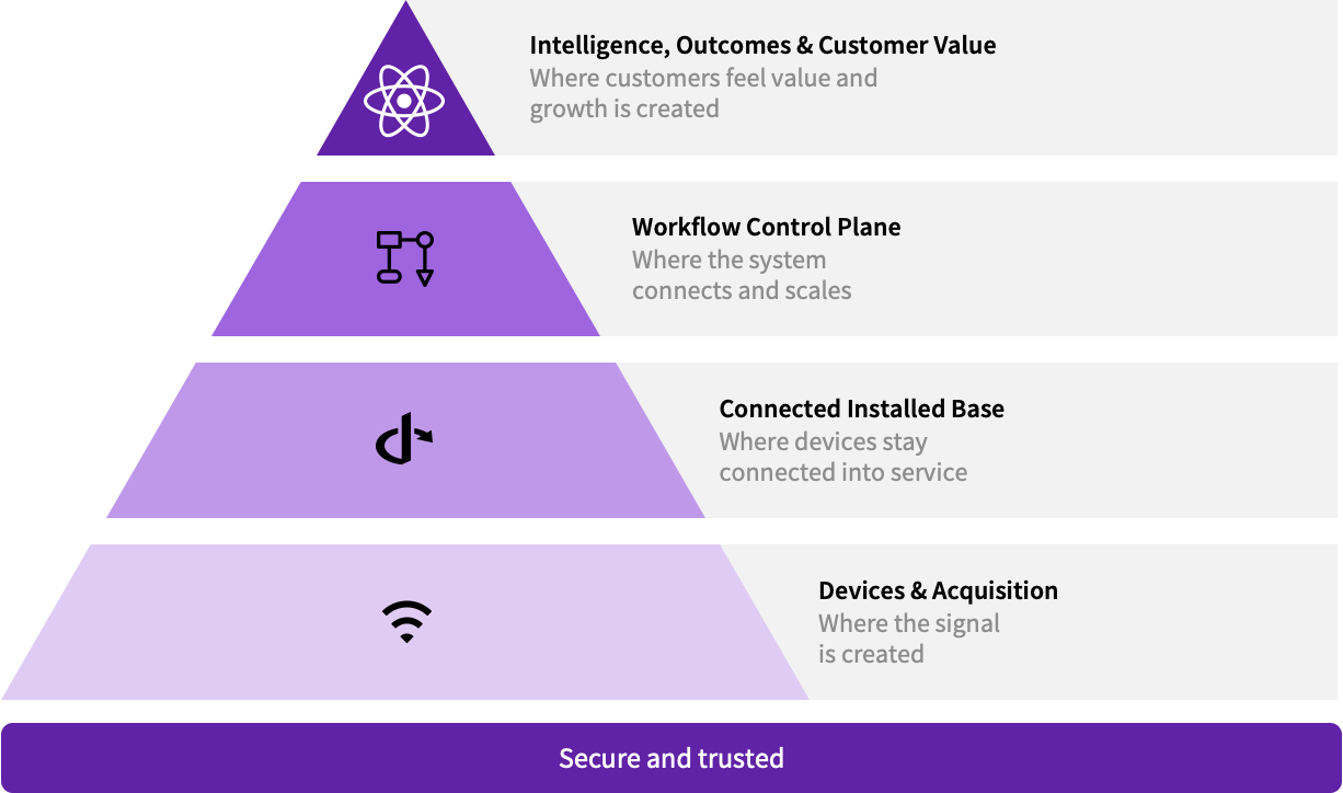
Caption: The architectural framework for a connected care pathway
The first layer is devices and acquisition. This is where images are created and where control over consistency, positioning, signal quality, device status, and fleet performance begins. In breast cancer care, the acquisition layer matters because the quality and consistency of the exam can affect downstream review and workflow.
Solutions like Invenia ABUS Premium are designed to support supplemental screening in women with dense breasts, consistent with their cleared indications for use. Use should remain consistent with applicable labeling, market authorization, and country availability.
The second layer is the connected installed base, where the system could connect and scale across the care pathway. This layer focuses on how data, tasks, and workflow information move across systems and teams. In the breast cancer journey, this may mean supporting coordination across screening, diagnostic follow-up, biopsy, and treatment referral in a connected flow. When implemented through appropriately authorized and integrated workflow solutions, this layer can help teams identify workflow gaps, coordinate handoffs, and support more timely follow-up coordination.
The third layer is the workflow coordination layer. This layer focuses on how breast imaging programs may manage capacity, coordinate resources, and support patient movement through the care pathway. These programs are not only managing interpretation. They are also managing staffing, room utilization, referrals, scheduling, callbacks, and patient access.
Supporting this work requires a shared digital foundation that can connect data, reduce integration burden, and make it easier to deploy new clinical and operational applications. CareIntellect is a family of clinical and operational applications that offers a cloud-first digital infrastructure, allowing healthcare providers to quickly and cost-effectively onboard new applications. Over time, this shared foundation is intended to support select AI-enabled applications across disease states and care settings, helping bring relevant information into existing workflows. Product functionality and availability vary by application and market.
The fourth layer is intelligence and workflow value. This is where a connected pathway could help ensure relevant information does not remain isolated within a single exam or system. Information should move with the patient through diagnostic evaluation, biopsy, referral, and follow-up. The goal is to reduce manual coordination, improve visibility across steps, and support a more coordinated pathway with fewer handoffs and less waiting.
Across these four layers, the aim is for AI to operate within bounded workflows. It should not replace clinician judgment. It should be designed to work within defined conditions, remain subject to human review, and support clinicians in delivering patient care.
When these layers are brought together, screening and breast cancer care could become more connected. Imaging can support image quality and consistency, while supplemental imaging for dense breast tissue can be incorporated into coordinated workflows. Operational tools can help care teams identify scheduling pressure and follow-up backlogs sooner. AI can support radiologist review while keeping the clinician in control. If cancer is diagnosed, the goal is for patient information to remain organized and accessible, reducing manual coordination and supporting more coordinated movement from screening to diagnosis and treatment referral.
Individual algorithms remain important. The next phase of healthcare AI, however, will be shaped by the ability to deploy them appropriately across connected care pathways. That means bringing devices, data, applications, workflows, and human review together in ways that can help reduce manual burden, support informed clinical review, and help patients move through the care journey more efficiently.